Amodo Design · Fish EEG
Fish EEG — Visually Evoked Potentials
A non-invasive way to read a fish's brain. A suction-cup electrode on the head picks up the EEG, a strobe flashes in front of the eye, and averaging across hundreds of flashes reveals the visually evoked potential (VEP) — a direct measure of whether the visual system is still responding. Watching the VEP fade as anaesthetic is delivered gives an objective marker of loss of sensibility, the question at the heart of humane stunning and slaughter.
The set-up
The fish rests in a shallow water tray inside a light-tight box. A tube in its mouth carries water dosed with anaesthetic over the gills, so the animal is ventilated and dosed at the same time. Every flash of the strobe is time-stamped alongside the EEG so the response can be averaged trial-by-trial.
The VEP fading under anaesthesia
Each frame is the moving average of the latest 200 flash trials (blue), against the first 200 trials of the session as a reference (grey). The sharp deflection just after the flash is the visual system responding. As the anaesthetic takes hold the response flattens and finally disappears.
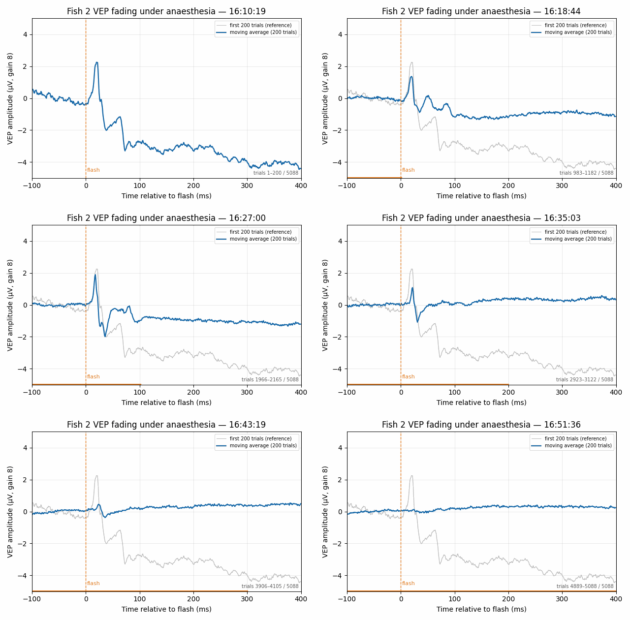
Fish 2, recorded 19th August 2026 · 5,088 flash trials · VEP amplitude in µV, time relative to flash in ms.
Why it matters
Non-invasive
The electrode is a suction cup on the skin. No implants, no surgery, and the fish can be returned to water once the recording ends.
Objective
Behavioural signs of insensibility in fish are ambiguous. A VEP is either there or it isn't, and the moment it goes can be measured to the trial.
Applicable at scale
The same measure can benchmark stunning methods, anaesthetic doses and recovery, giving processors and regulators a common yardstick for fish welfare.
Non-invasive, trial-synchronised, quantified loss of sensibility.
gill irrigation with dosed anaesthetic · 200-trial moving average